Virchow 2.0: From intervention to prevention
Bridging the gap between basic research and clinical practice and bringing cell-based medicine to the clinic – these are the aims of the research network “Virchow 2.0 – Berlin Brandenburg innovation cluster for cell-based medicine.” October 1 marks the official start of the conception phase of the network, which is one of the 15 finalists in the second round of competition in the Future Clusters Initiative (Clusters4Future) of the German Federal Ministry of Education and Research (BMBF) and the only finalist from Berlin. The core partners of Virchow 2.0 are the Max Delbrück Center for Molecular Medicine in the Helmholtz Association (MDC), the Charité – Universitätsmedizin Berlin, the Berlin Institute of Health at Charité (BIH), the Zuse Institute Berlin (ZIB), and the Berlin Institute for the Foundations of Learning and Data (BIFOLD), a Berlin research network that develops applications for big data and machine learning.
Now, some 160 years after Virchow, we have technologies that enable us to create a cell-based medicine.
Virchow 2.0 stands for tradition and the cluster’s vision for the future. In the 1850s Rudolf Virchow laid the foundation of cellular pathology, which posits that diseases are based on disorders of the cells and their functions. Scientists have further developed this approach, which was revolutionary at the time. “Now, some 160 years after Virchow, we have technologies that enable us to create a cell-based medicine,” explains Professor Nikolaus Rajewsky, who as Scientific Director of the Berlin Institute for Medical Systems Biology (BIMSB) at the MDC is coordinating the initiative. Co-spokesperson is Professor Angelika Eggert, head of the Department of Pediatric Oncology and Hematology at Charité – Universitätsmedizin Berlin. “These technologies include groundbreaking single cell and imaging methods, which we will combine with artificial intelligence and personalized disease models such as organoids,” adds Rajewsky.
“Rudolf Virchow would be thrilled”
“These methods essentially take us back to the roots,” says Professor Frederick Klauschen, head of the Systems Pathology Group at the Institute of Pathology at Charité – Universitätsmedizin Berlin, Director of the Institute of Pathology at Ludwig-Maximilians-Universität München, member of the Berlin AI center BIFOLD, and co-coordinator of the MSTARS research consortium. The consortium’s mission is to further develop mass spectrometry as a tool for patient care and to use the technology to get to the bottom of what causes therapy resistance. “Molecular studies on fragmented tissue are more of a stopgap than a solution, as we have so far lacked the tools needed to study single cells,” says Klauschen. “I wonder what Virchow would have said about this – after all, pathologies have always been about single cells.”
“Rudolf Virchow would be thrilled if he were alive today,” Rajewsky says with conviction, explaining: “We will be able to use the first cellular changes to diagnose diseases, predict possible disease trajectories, and steer the molecular networks away from the emerging disease and back toward a healthy equilibrium. It will also enable us to identify new targets for therapeutic agents and cell therapies.”
An all-around transformation of research and clinical practice
The research being done by the initiative’s core partners is already showing what tomorrow’s cell-based medicine may look like. “Cell-based medicine doesn’t just mean producing cell products to treat patients; it also means we can identify cell types that might cause a particular disease or bring about a healthy equilibrium,” says Professor Birgit Sawitzki. “If we understand why they differentiate in one direction or the other, we may also find out how to switch them off or increase their number.” She and her team at the BIH are investigating the molecular mechanisms that underlie immune cell activation and inhibition. Their studies focus on T cells, which recognize and fight foreign structures like viruses or bacteria. T cells also stimulate the formation of B cells, which in turn produce antibodies to attack the invaders. To analyze the cells, Sawitzki combines single-cell RNA sequencing with multiparameter flow cytometry and mass spectrometry. This has led her to discover that certain T cells accumulate in the lung tissue of COVID-19 patients and release molecules that damage lung cells. So on the one hand, the scientists have identified biomarkers that can help to predict with great accuracy the severity of the COVID-19 disease, while on the other hand they seek to find out how they can suppress the signaling pathway that triggers the formation of these tissue-damaging T cells.
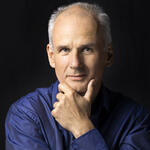
Prof. Dr. Heyo K. Kroemer, Chief Executive Officer of the Charité
Dr. Annette Künkele of Charité is also doing research on T cells. She and her team at the Department of Pediatric Oncology and Hematology are working to develop new immunotherapies against neuroblastoma, a cancer of the nervous system that mainly affects young children up to six years of age. They are using a technique called CAR T-cell therapy that involves taking T cells from the patient and equipping them with a chimeric antigen receptor (CAR) in the laboratory. Armed with these receptors, the T cells are returned to the patient’s body, where they are able to find and destroy cancer cells. CAR T-cell therapy has already been used with great success in the treatment of leukemias. Yet solid tumors like neuroblastoma are better able to fend off attacks by T cells. “We want to uncover and disable those defense mechanisms that currently prevent a cure,” says Künkele. In her research group, she is collaborating with national and international partners on studies that investigate various CAR constructs and how tumor cells, their environment, and surrounding blood vessels influence the success of CAR T-cell therapy.
Further research examples
- 3D maps of gene activity
-
-
Together with Dr. Nikos Karaiskos from his research group at BIMSB, Nikolaus Rajewsky has succeeded in creating a kind of spatial map of gene expression for individual cells in different tissue types. The scientists used single-cell sequencing data to develop a mathematical model that can calculate the spatial pattern of gene expression for the entire genome, thus enabling them to precisely track whether or not a particular gene is active in the cells. “The three-dimensional measurement of gene expression is one of the driving forces in single-cell biology,” Rajewsky says with conviction. “To understand exactly what happens in human cells during disease progression, we not only need to decipher the activity of the genome in individual cells, but we also and more importantly need to track it spatially within an organ.” This will allow us to diagnose the disease accurately and select the optimal therapy.
- New generation of organoids accelerates drug development
-
-
Organoids are mini-organs in a petri dish. They are grown from human cells and mimic the tissue architecture, cellular diversity, and genetic signature of a real organ. This means they can serve as patient-specific disease models that reveal how cells interact and respond to substances. Yet until now organoids have for the most part been non-scalable and difficult to reproduce, making them costly to use in research and thus limiting their viability. Dr. Jakob Metzger, who heads the Quantitative Stem Cell Biology Lab at the MDC’s BIMSB, is working with his team to create neuronal organoids that can grow in 96-well plates and thus be used for drug screening. “By combining reproducible organoids with high-throughput methods, we’re able to generate data that can be used for machine learning,” says Metzger. This creates great opportunities for drug therapy development – which has so far been a very lengthy process for which only very few candidates are ever considered.
- Organs-on-chips mimic processes in the human organism
-
-
Professor Sarah Hedtrich studies atopic diseases at the BIH. These include asthma, neurodermatitis, and allergic rhinitis with conjunctivitis, as well as hay fever and house dust mite allergy. To understand the mechanisms behind these diseases, she is building three-dimensional human skin models from the cells of both diseased patients and healthy donors. The scientist is also using so-called tissue engineering to develop bronchial epithelium models. “Our goal is to create an organ-on-a-chip that contains blood vessel-like structures, which even use special pumps to simulate blood pressure,” she says. “Such models are very good at emulating processes in the human organism.” Hedtrich is convinced that this applies not only to diseases of the skin and lung epithelium: “Much of the failure in drug development is due to the fact that results obtained in mice or rats cannot be translated to humans. Using complex human models can help improve translation, and would therefore bring great advantages not only in terms of animal welfare.”
- Explainable AI
-
-
Pathologists usually determine if a suspicious lump is cancerous or not. In Germany there are 1,200 pathologists and 500,000 new cancer cases every year. The use of digital assistant systems can help avoid mistakes. “Humans are not as good at determining what percentage of tissue is affected by cancer or what percentage of tumor cells contain a certain therapeutically relevant receptor. Our ‘digital colleague’ is both faster and more precise when it comes to quantitative analysis,” explains Frederick Klauschen. His team from the Systems Pathology Group at Charité’s Institute of Pathology and colleagues from TU Berlin have joined forces to develop a digital imaging analysis system that uses artificial intelligence (AI) to evaluate microscopic images. The system can also show us how the AI makes its decisions. The software creates so-called heat maps, which show precisely which cells or image areas were decisive for the algorithm’s classification of the tissue as cancerous or non-cancerous. This allows the pathologists to assess whether the AI analysis is plausible. The technology also opens up entirely new possibilities for research. If, for example, one trains the AI with the positive and negative courses of a particular therapy, the resulting heat maps could enable pathologists to discover biomarkers that predict therapeutic success. This “explainable AI” technology is being further developed in the young spin-off company Aignostics.
Kroemer: We’re taking prevention to a new level
“The importance of this new emerging branch of science for medicine is immense,” says Eggert. Within the Virchow 2.0 cluster she ensures the link between basic research and clinic practice: “With the help of single-cell methods, we can define drug targets very precisely. Personalized disease models enable individually tailored drug testing. They also allow us to test in advance which treatment is most effective.”
Professor Heyo Kroemer, Chief Executive Officer of Charité, is also convinced of the potential of cell-based medicine: “Medicine today is mainly about therapeutic intervention. Yet with the help of new technologies, we will be able to prevent diseases from occurring in the first place. This will take prevention to a whole new level.”
Keeping an eye on the market
The participating experts from systems biology, medicine, biotechnology, physics, and computer science/artificial intelligence want to team up with partners from local and national industry to build an AI-driven biomedical innovation ecosystem. The cluster also aims to create a favorable environment for spin-off businesses and a support platform to involve established companies in order to pave the way for the translation of scientific discoveries into clinical practice.
Virchow 2.0 offers us the opportunity to connect these different players and look at diseases from a completely new perspective using single-cell methods.
“We are not starting from scratch,” stresses Thomas Gazlig, who heads Charité BIH Innovation, the joint technology transfer office of the BIH and Charité that supports prospective company founders. Both the MDC and the BIH and Charité have technology transfer divisions that help scientists commercialize discoveries and translate them into practice as quickly as possible. These divisions offer coaching and mentoring programs while also helping with funding applications and the search for development and commercialization partners. Gazlig sees solid science as the foundation of Virchow 2.0, pointing out that “it’s our job to find and promote disruptive innovations, but we have to keep an eye on the market to become true game changers.”
“There’s huge potential to make this happen in Berlin-Brandenburg,” says Dr. Ashley Sanders, who as head of the Genome Instability and Somatic Mosaicism Lab at the MDC is part of a research collaboration between the BIH, Charité and the MDC that aims to advance single-cell technologies. She adds that the area is home to a constellation of regional players from basic research, clinical medicine, and application-oriented R&D that is not only unique in Germany but also includes global leaders in the relevant technology, data science, and medical fields. “Virchow 2.0 offers us the opportunity to connect these different players and look at diseases from a completely new perspective using single-cell methods,” says Sanders. Dr. Kai Uwe Bindseil, head of the Division of Health Economy, Industry, and Infrastructure at Berlin Partner for Business and Technology, assesses Virchow 2.0 from the perspective of the local economy as follows: “The Berlin-Brandenburg region has everything a successful cluster needs: scientific excellence, a strong will to collaborate, a high international reputation, and political support. Successful start-ups like T-knife, Ada Health, and Care Syntax have raised a total of €300 million in venture capital in recent months – proof that the region is very attractive for investors.”
The Clusters4Future competition
Under the umbrella of the High-Tech Strategy 2025, the German Federal Ministry of Education and Research (BMBF) seeks to strengthen knowledge and technology transfer through the open-topic competition Clusters4Future. It aims to foster optimal collaboration in a region among different actors from science, academia, industry and society. The German government plans to provide up to €450 million in total funding over the next 10 years for the Future Clusters.
In the second round of the competition, scientists were called on to submit proposals from their chosen field, such as robotics, energy or biomedicine, for the creation of regional innovation networks – the so-called Future Clusters. An independent jury of experts has now selected the best 15 of the 117 cluster ideas to participate in the conception phase. The BMBF is supporting this six-month phase with up to €250,000.
The individual cells isolated from the tissue are shuttled into miniaturized chips for analysis.
In the conception phase, which began with the meeting in Berlin, the participants are developing the cluster strategy including the various research and development projects and innovation supporting activities for the initial implementation phase. There is great interest in joining the cluster on the part of science and industry: At the kick-off alone, 25 researchers and innovators shared their project ideas for making the vision for cell-based medicine come true. Some 20 companies already support the initiative. “We also want to work together with patient organizations,” stresses Eggert, “because no one knows the needs of people suffering from disease better than they do.”
In mid-2022, up to seven Future Clusters will be selected based on the vote of an independent jury of experts. These clusters will be able to implement their concepts for up to nine years. Up to €5 million in funding is available per cluster and year.
Text: Jana Ehrhardt-Joswig
Further information
- Presse release: A leap into the future with Virchow
- Future Clusters Initiative of the BMBF (German only)
- Medical Systems Biology: The Berlin Institute for Medical Systems Biology
- Single-cell analysis at the MDC
- Single cell approaches for personalized medicine
- The visionary – portrait of Professor Nikolaus Rajewsky
- Berlin should establish a cell hospital (German only)
- LifeTime Initiative
- BIFOLD
- Zuse Institute Berlin


